
![THE HERALD STUDENT REPORTERS [US]](/assets/images/logo_student_us.png)
![THE HERALD STUDENT REPORTERS [Canada]](/assets/images/logo_student_ca.png)

Trained AI may become better at Cancer diagnosis than pathologists
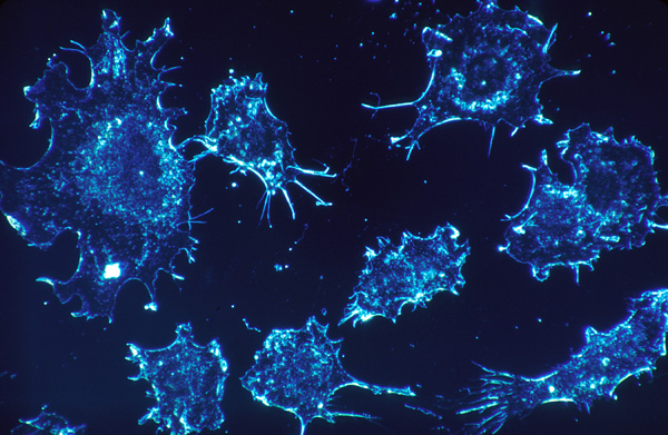
[Cancer cells. Photo Credit to Raw Pixel]
Researchers led by University of Pennsylvania pathologist Zhi Huang have developed an artificial intelligence system trained to emulate how human pathologists search tissue slides, significantly improving cancer-spotting accuracy and pointing toward a new collaborative role for AI in medical diagnostics.
In clinical practice, confirming cancer begins when suspicious screening results or physical symptoms prompt a tissue biopsy.
Pathologists then examine microscopic sections, analyzing cellular morphology, tissue architecture, and invasion patterns to compile a diagnostic report that guides treatment.
Although computational tools have increasingly entered this field, conventional pathology AI models typically evaluate fixed-size image patches or preselected static regions.
Because a single digital slide contains billions of pixels while evidence of cancer may occupy only a tiny fraction, traditional patch-based systems can easily miss subtle abnormalities or generate excessive computational noise.
To overcome these limitations, Huang’s team designed a system called Pathology-o3, inspired by the dynamic search strategies of human experts.
Rather than analyzing isolated image tiles, human pathologists approach a slide much like a search-and-rescue helicopter: they perform a rapid, low-magnification sweep of the visual landscape before zooming in on specific “red flags” for closer examination.
To capture this active decision-making process, researchers developed a software tool that recorded live diagnostic sessions from eight expert pathologists.
The software logged every interaction, including pans across the tissue and magnification shifts between low-power overviews (5x to 10x) and high-power detail views (40x).
Because raw human navigation is inherently imprecise—filled with accidental mouse drifts, overshoots, and minor manual adjustments—the researchers systematically filtered out incidental movements.
They isolated deliberate attention patterns and validated them against eye-tracking data to ensure the software captured exactly where the experts focused.
Additionally, Vision-Language Models (VLMs) drafted concise visual explanations for why flagged regions appeared suspicious—such as highlighting atypical cell clusters indicative of metastasis—which human pathologists then reviewed, edited, or approved.
By structuring these refined actions into a training dataset called "Pathology-CoT" (Visual Chain-of-Thought), the team trained Pathology-o3's internal behavior predictor to automatically anticipate where an expert would look and prioritize these critical regions for high-resolution evaluation.
In tests involving colorectal cancer lymph node slides, Pathology-o3 correctly identified 100% of cancer-positive slides, surpassing OpenAI’s general-purpose o3 model, which achieved an 87.5% detection rate.
Pathology-o3 also registered a 15.5% false-positive rate, a substantial improvement over o3’s 53.3% false-positive rate.
When evaluated on an independent dataset from an unfamiliar institution, Pathology-o3 maintained a high positive detection rate of 97.6%, though its false-positive rate rose to 37.1%—underscoring how variations in tissue staining and digital scanner calibration continue to challenge model generalization across different laboratories.
Despite these encouraging results, significant clinical and operational concerns remain.
Elevated false-positive rates can increase pathologist workloads by creating unnecessary visual alerts.
Furthermore, current AI models evaluate individual slides in isolation, whereas comprehensive clinical diagnoses rely on synthesizing multiple stained tissue sections, molecular testing, and broader patient histories.
Broader challenges surround the "black box" nature of deep-learning decisions, data privacy, and legal liability when an algorithm errs also persists.
Consequently, experts emphasize that tools like Pathology-o3 are intended to serve as intelligent prescreening assistants rather than autonomous replacements for human pathologists.
The ultimate test for this technology will be prospective, multi-center clinical trials that measure whether pathologists working alongside AI assistants actually diagnose more cancers, make fewer errors, and manage workloads more efficiently in daily practice.

- Seungmin Shin / Grade 12
- North London Collegiate School Jeju